


UCLA researchers streamline skin tissue analysis with virtual staining technique
(Katie Frei/Daily Bruin)

By Aditi Kumar
Jan. 17, 2022 4:13 p.m.
UCLA researchers have developed a virtual, non-invasive method of visualizing skin tissue that could transform how skin diseases are diagnosed.
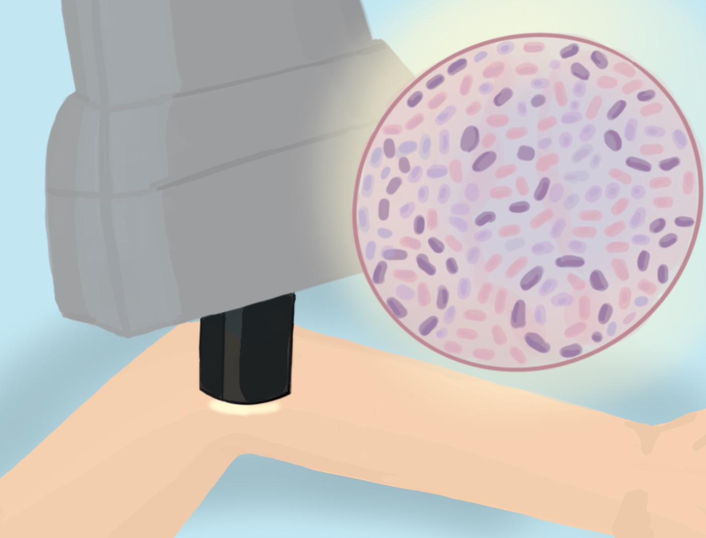
In a Nov. 18 study, UCLA researchers described how artificial intelligence could be used to transform a black-and-white image of the skin obtained by a non-invasive microscope into a colorized, highly-detailed image, similar to one obtained through a biopsy.
A traditional biopsy requires that a skin sample be removed from a patient’s body, separated into layers and stained with dyes in order to visualize the details within cells, said Aydogan Ozcan, a distinguished professor in electrical and computer engineering and co-author of the study. Depending on the tissue and stain combination, the staining process can take up to a day. For difficult-to-perform stains, there can be variability in the results, he added.
Jingxi Li, a co-author of the study and doctoral student in electrical and computer engineering, said stains traditionally used for tissue samples, such as H&E, can lead to deformation of the sample.
“Our experience working with some of the hospitals in (the) Los Angeles area (was that) 30% of the time the staining fails (for difficult-to-perform stains),” Ozcan said.
Dr. Gene Rubinstein, volunteer clinical faculty in the division of dermatology at the David Geffen School of Medicine and a co-author of the study, said that the process of taking a skin biopsy had remained the same for about 100 years until scientists developed the reflective confocal microscope, which could produce black-and-white images of the skin without a biopsy.
The person operating the RCM would only need to place it on the patient’s skin to capture an image below the surface, Li said.
The RCM is FDA approved, but the images it produces are difficult to interpret, preventing it from being used throughout clinics.
“The issue with that technique has been that it’s difficult to interpret for people who are not used to interpreting it,“ said Rubinstein, who is also CEO and medical director of the Dermatology and Laser Centre in Studio City. “And that was the reason why we decided on this collaboration between myself and UCLA and Dr. Ozcan.”
Their new method builds on the existing technology of the RCM, replacing chemical staining with a computerized version called virtual staining.
Grayscale images of skin tissue obtained with the microscope are entered into a series of computer algorithms known as an artificial neural network, Ozcan said. This network then produces colorful, detailed images that appear as though they were actually stained.
“Think of it like a black-and-white movie,” Ozcan said. “You can take that black and white movie and … through a deep neural network, represent how it’s supposed to be if it were to be shot in color.”
[Related: Researchers at UCLA develop computer-free technology for deciphering holograms]
Li, Ozcan and Rubinstein believe that the benefits of this method would be widespread.
According to Ozcan, their method could help scientists in parts of the world that may lack access to the proper tools to visualize human cells and tissue and allow patients to diagnose patients remotely through telemedicine. It could also help conserve water, since large amounts of wastewater are generated during the staining process.
Rubinstein said their method could prevent “loss to follow-up” – where the patient does not return for follow-up appointments for multiple reasons – by diagnosing diseases immediately and removing diseased tissue if necessary.
Li added that the method could eliminate many costs that add up for a traditional biopsy.
The researchers are still hoping to make changes to the method before it can become an established part of clinical practice.
The RCM is still not small enough for dermatologists to conveniently use, Rubinstein and Li said. Future research should aim to improve the quality of the virtually-stained images and the speed at which they are produced, they added.
Nevertheless, Ozcan said he believes this method is the first of its kind and a breakthrough for diagnosing skin diseases.
“It’s a challenging project and I think it’s a milestone,” Ozcan said.

