


Virtual reality researchers model heartbeats of zebrafish to use in medicine
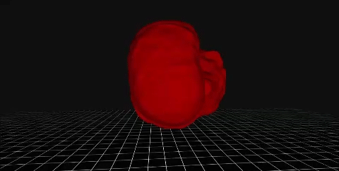
(Courtesy of JCI Insight) A visualization of a zebrafish’s heart from a previous paper the lab published.

By Raymond Le
Sept. 27, 2018 2:09 a.m.
This post was updated Oct. 2 at 4:58 p.m.
UCLA researchers developed a virtual reality visualization of zebrafish hearts for use in medical treatment and surgical training.
In a paper published Aug. 15, the Cardiovascular Engineering & Light Sheet Imaging Lab under the Department of Bioengineering created a new computer program that can generate a 4-D representation of a zebrafish’s beating heart, using time as the fourth dimension.
Rene Packard, an assistant professor at the David Geffen School of Medicine who works at the lab, said the researchers used zebrafish because they regenerate organs quickly, which allows the researchers to conduct multiple experiments and collect measurements in a short time period.
Arash Abiri, a UC Irvine graduate and researcher in the lab, said he noticed most models of beating hearts in prior visualizations were choppy and unrealistic.
“It didn’t have the fluid movements you’d want a beating heart to have,” Abiri said. “This inspired me to not lose the integrity of the motion when reconstructing the models.”
The researchers used a light sheet fluorescence microscope to obtain thousands of images of 2 micrometer-thick slices of zebrafish hearts.
“We built our own optical microscope that allows for many lenses and lasers to focus on the organ of interest, and through this complex array you can generate an image,” Packard said.
Previously, researchers had to manually outline each section of the heart, such as the atrium and ventricles, on 4,000 different images.
“(The images) cannot be put on a computer, as it cannot tell which part is a ventricle or atrium,” said Yichen Ding, a postdoctoral researcher in the lab.
With the new computer program, researchers only had to outline 86 images while the computer automatically outlined the rest, allowing for a fluid and dynamic visualization.
Abiri said this simulation can be used with Google Cardboard, an inexpensive virtual reality headset.
This allows doctors or researchers to visualize the interactions between parts of the heart and see the interactions’ effects on the heart’s structure and development, Packard said.
“VR allows you to see the heart from the inside and follow structures of interest, such as seeing a cell group’s contribution to cardiac development,” Packard said. “You can go virtually inside the heart and see the interaction of groups of cells, and how cardiac processes affect heart mechanics and development.”
Abiri said he hopes this research technique will be applied to other organs in the body.
“It can be applied to a heart attack – a portion of (cardiac) tissue may die, so if you get a CT image of that person’s heart and visualize it you can view in VR the different parts of the heart moving and see that helping with medical treatment and surgical planning,” Abiri said.

