


UCLA researchers develop camera, technique to find cancerous tissue
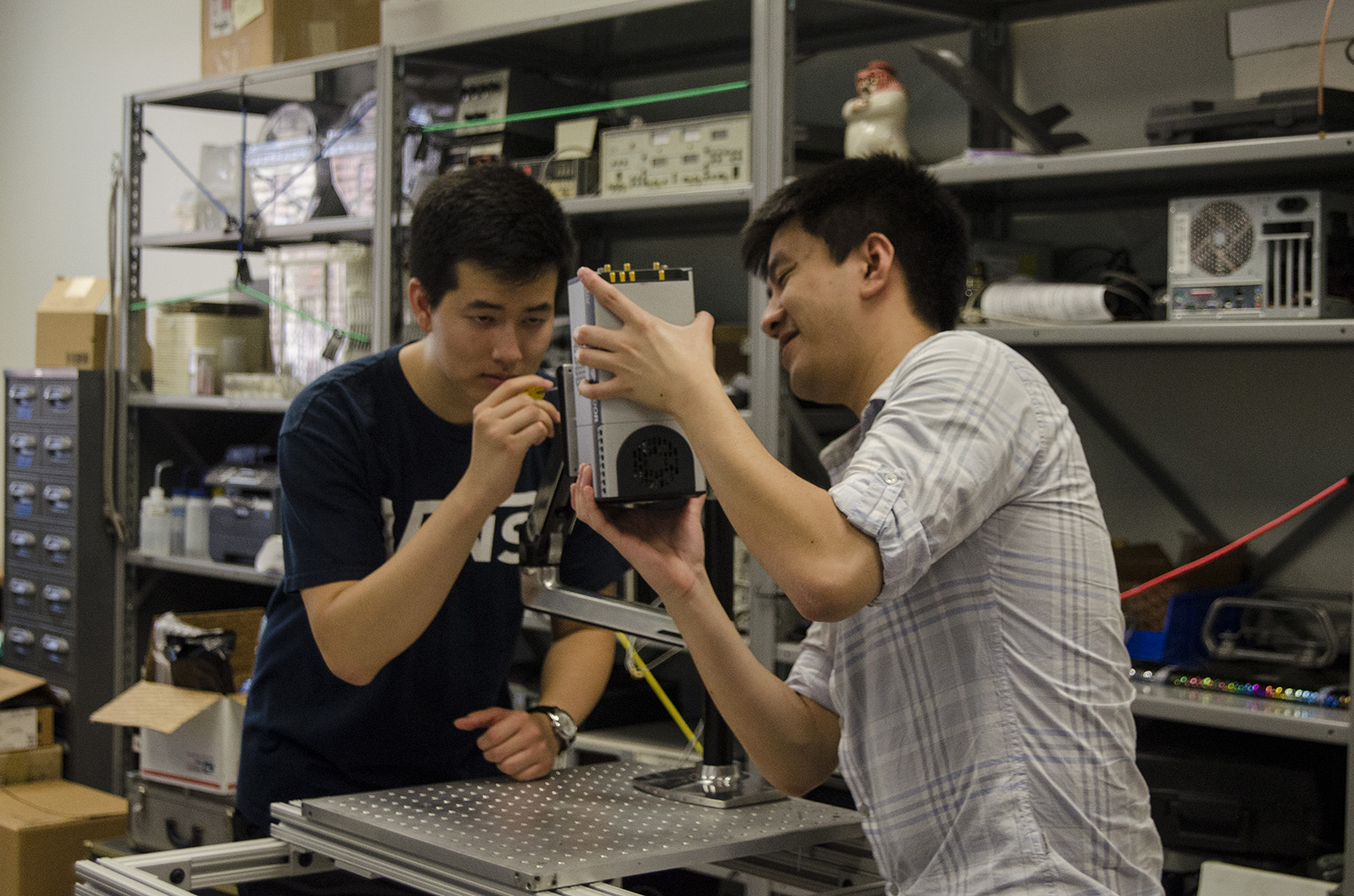
Harrison Cheng, researcher and graduate student in engineering, holds the camera steady as Zachary Tom, researcher and second-year bioengineering student, screws it to a metal beam. (Miriam Bribiesca/Daily Bruin senior staff)
By Daily Bruin Staff
Oct. 8, 2015 4:07 a.m.
With steady arms, Harrison Cheng raised a camera up to a metal beam while his colleague inched slowly toward it, securing it with a screwdriver. The two researchers carefully assembled a camera arm that would hang over surgeries to take photographs of cancerous tissue.
Researchers will begin to use the charge-coupled device camera in trials at the Ronald Reagan UCLA Medical Center on Monday, said Zachary Tom, a researcher and second-year bioengineering student. The trials are a part of bioengineering professor Warren Grundfest’s long-term research study that aims to use a novel quasi-real-time fluorescence lifetime imaging technique to better distinguish between cancerous and noncancerous tissue before surgeries.
General fluorescence imaging is a technique that uses an external source, such as a laser, to shine a light on the tissue. The light is absorbed by the tissue and emits lower energy that is captured by a camera, which will then create an image of the tissue on a screen, said Aidan Pearigen, a researcher and third-year bioengineering student.
Surgeons currently require at least 30 minutes to transport, process and analyze tissue samples to determine whether a tissue sample contains cancerous cells. Quasi-real-time fluorescence lifetime imaging allows surgeons to determine the cancerous cells within a minute, said Cheng, a research assistant and graduate student in bioengineering. Surgeons can then quickly determine the location of the malignant tissue and minimize healthy tissue removal.
For example, doctors must minimize healthy brain tissue removal in patients with brain tumors because unnecessary extractions could lead to the deterioration of different bodily functions, Pearigen said.
Surgeries using conventional fluorescence lifetime imaging are time consuming because this technique uses complex formulas to categorize different types of tissue, Cheng added. Quasi-real-time fluorescence imaging uses simple mathematical algorithms to minimize the time it takes to classify the samples.
Pearigen said he began working in Grundfest’s lab about a year ago because he wanted to explore the field of bioengineering. He added he thinks the application of fluorescence lifetime imaging is critical in helping patients.
“Sometimes with cancer, if the doctor does not remove all of the necessary tissue, the cancer cells can regrow and have horrible consequences,” he said.
Cheng said he became involved in the research because of his interest in optical imaging.
“I’m interested in trying to find optical imaging techniques that can do real-time (cancer) detection, like video streaming,” he said.
Because the project combined mechanical and electrical engineering as well as computer science concepts, the researchers had to learn crucial information about the other fields to assist in the research and create the device, Cheng said.
Tom added the device will be tested in the coming months at the Ronald Reagan UCLA Medical Center and St. John’s Health Center, but will not be used commercially for several years.
“I feel like I am making a difference and working towards improving medicine for the future,” Tom said. “Our research is going really bright, as in the next few months, we can get the system ready.”