


New laser microscope technology allows scientists to see neuron activity of the entire brain

By Nicole Arulanantham
Jan. 18, 2011 1:10 a.m.
Adrian Cheng was an undergraduate student studying physics and math at UCLA just eight years ago, uncertain of how he could contribute to the scientific field.
Today, Cheng is the inventor of a microscope that allows scientists to view the workings of the brain as a whole.
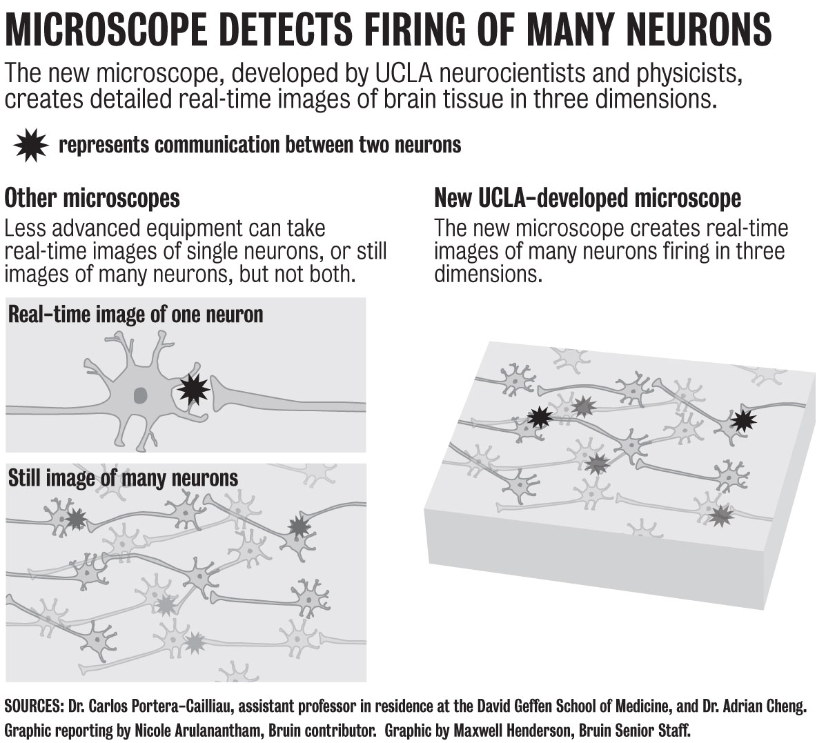
The microscope will allow researchers to study the patterns of signals sent from neuron to neuron, said Carlos Portera-Cailliau, an assistant professor in residence at the David Geffen School of Medicine.
He compared viewing neuron activity in the brain to observing the proceedings of an ant colony.
Prior to the invention of the new microscope, scientists studying the brain were only able to view the equivalent of a single ant or a still image of the entire colony. Now they will be able to see what amounts to the activity of all the ants in the colony at once.
Previous microscopes used a single laser beam to scan brain tissue and form images of the cells.
“But laser beams can only go so fast,” Portera-Cailliau said. These beams could not return data quickly enough to capture an image of a firing neuron, a process that occurs in about a millisecond.
Scientists then began using multiple laser beams, which made the process of collecting images much faster. The problem with this method was that the brain caused light particles from the beams to scatter.
When the particles scattered, it was impossible to tell which beams were in which area of the brain. While this technology could be used at the surface of the brain, it did not work when trying to image deep into the brain.
To get around this, Cheng suggested that they send out the laser beams one at a time with a small delay in between. This allowed the detector to determine which area each beam was scanning, which returned detailed images of individual neurons firing in the brain.
While the study of the brain is a primary focus of neurologists, the technology behind the microscope’s design also required the expertise of physicists. Research has been in progress for about 10 years, but Cheng said the partnership between the disciplines was only formed about four years ago.
UCLA professor Katsushi Arisaka, who was Cheng’s adviser, said he hopes they can commercialize the product and bring it to other campuses and institutions.
“It’s exciting to realize that you’re going to contribute to the research instead of simply being part of the audience,” Cheng said. “We really hope to continue to develop the technology so researchers at large can use it.”


